NanoTools Facility Instrumentation
Bruker D8 Discovery 2D X-ray Diffractometer
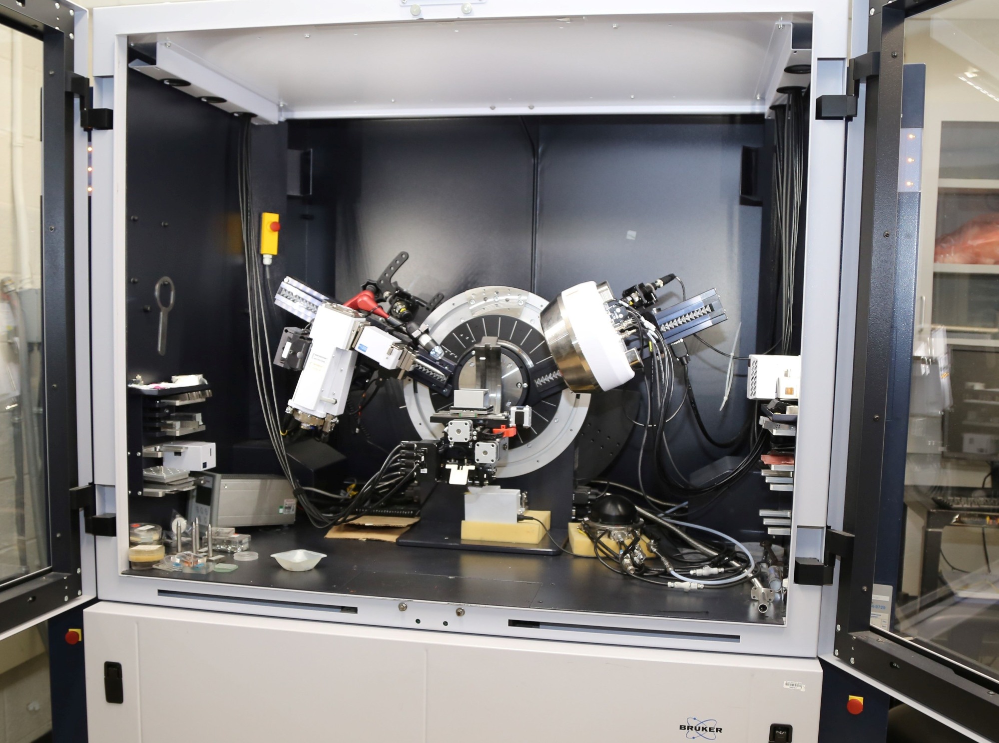
This Bruker D8 Discover X-ray Diffraction System has a twist tube for line and point focus applications. A Vantec 500 2D area detector and a LinxEye (0D, 1D) detector for analysis. The system is suitable for texture, stress, and micro-x-ray diffraction as well as for orientation and epitaxy studies. The instrument has in-situ capabilities for studies at low/high temperature and under vacuum to study properties as a function of time, temperature, gas environment or vacuum. A capillary spinner stage is also available for investigations of air sensible materials with the sample prepared in a capillary glass.
Bruker D8 Discovery High Resolution X-ray Diffractometer
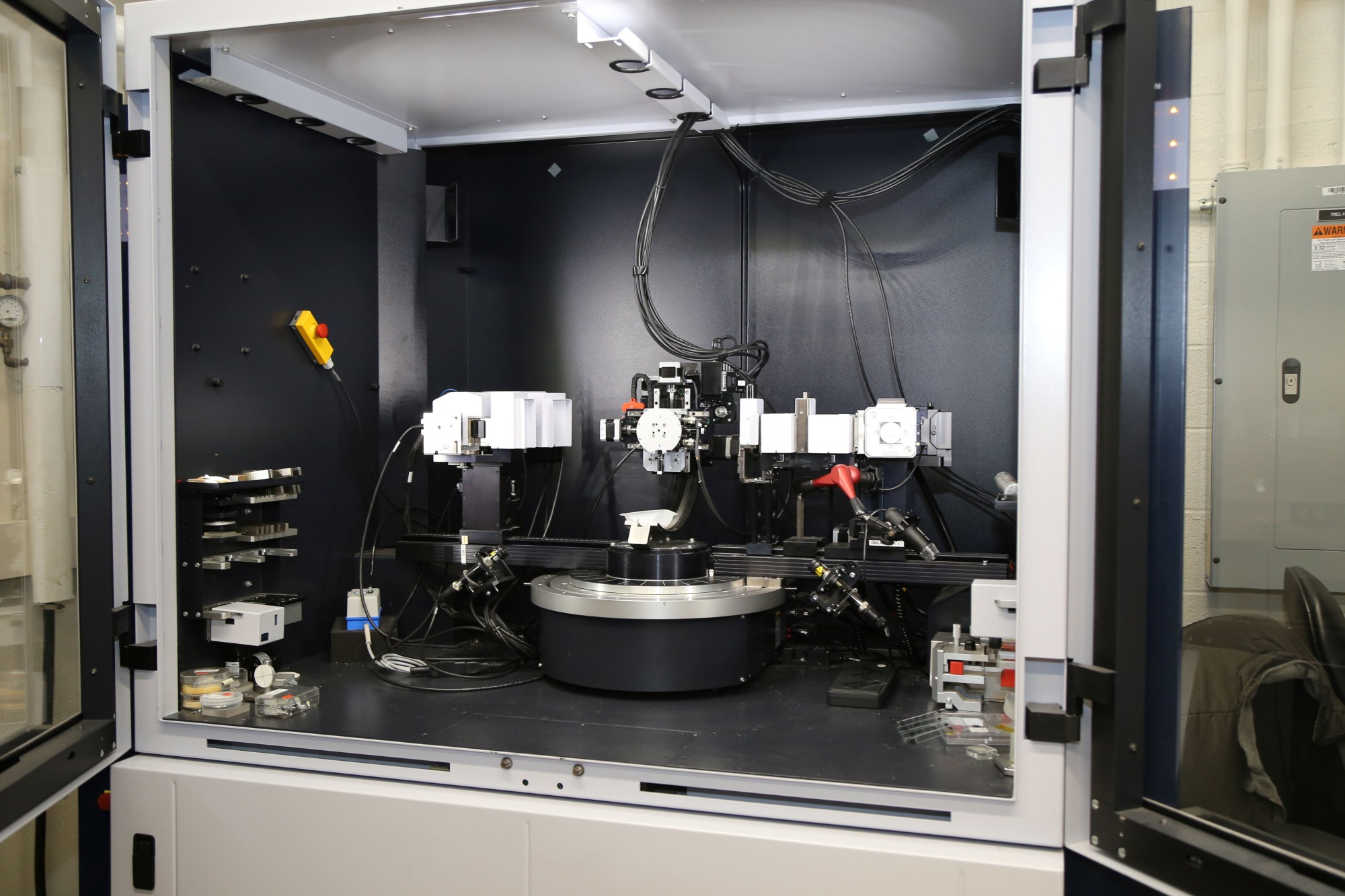
This Bruker D8 Discover X-ray Diffraction System includes a line source configuration system with a LinxEye (0D, 1D) detector and a Scintillator detector. The system is suitable to determine thickness, crystallographic structure and strain in epitaxially thin films as well as high resolution analysis of bulk materials. X-ray reflectometry (XRR), high resolution XRD, grazing incidence diffraction (GI-XRD), in-plane GID (IGID) and reciprocal space mapping (RSM) are some techniques that can be performed with this configuration. High resolution is obtained using parallel beam optics: Goebel mirror, monochromators, soller slits and analyzer crystals.
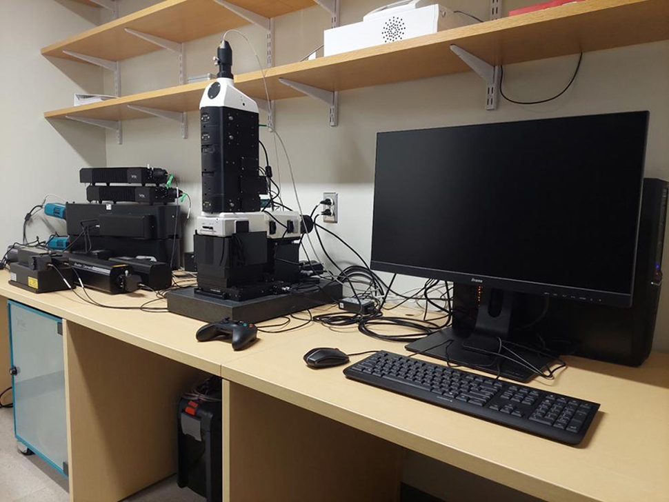
Asylum MFP-3D Origin Atomic Force Microscope
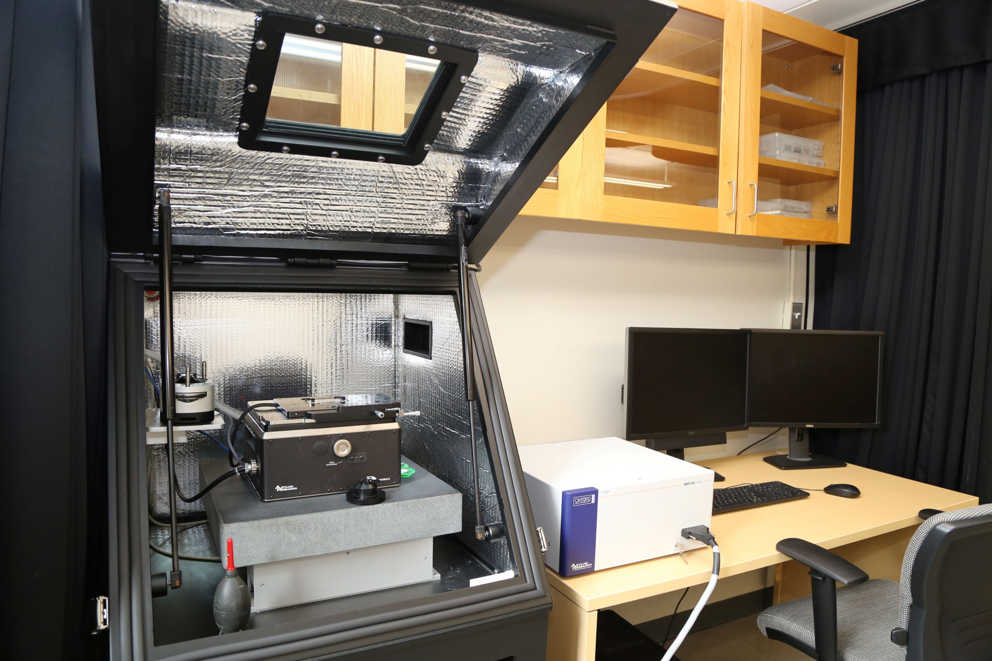
The Asylum MFP-3D Origin atomic force microscope (AFM) is a multi-mode instrument. It is equipped with a closed loop operation in all three axes with high precision, resolution and low noise operation. The closed loop Z scanner eliminates hysteresis. The AFM provides a 90 µm X and Y range (< 0.5 nm noise) and 15 µm Z range (Z sensor, 0.25 nm noise) and detector noise <15 pm. It also includes Inverted optical lever design that eliminates interference artifacts and a low-coherence infrared (860 nm) super-luminescent diode (SLD) light source (class 1M). Applications include dual AC resonance tracking (DART), electric force microscopy (EFM), force mapping mode, lateral force microscopy (LFM), magnetic force microscopy (MFM), kelvin probe force microscopy (KPFM), piezo-response force microscopy (PFM), and conductive AFM (C-AFM) with ORCA.
Bruker EMX Premium-X Electron Paramagnetic Resonance (EPR) Spectrometer
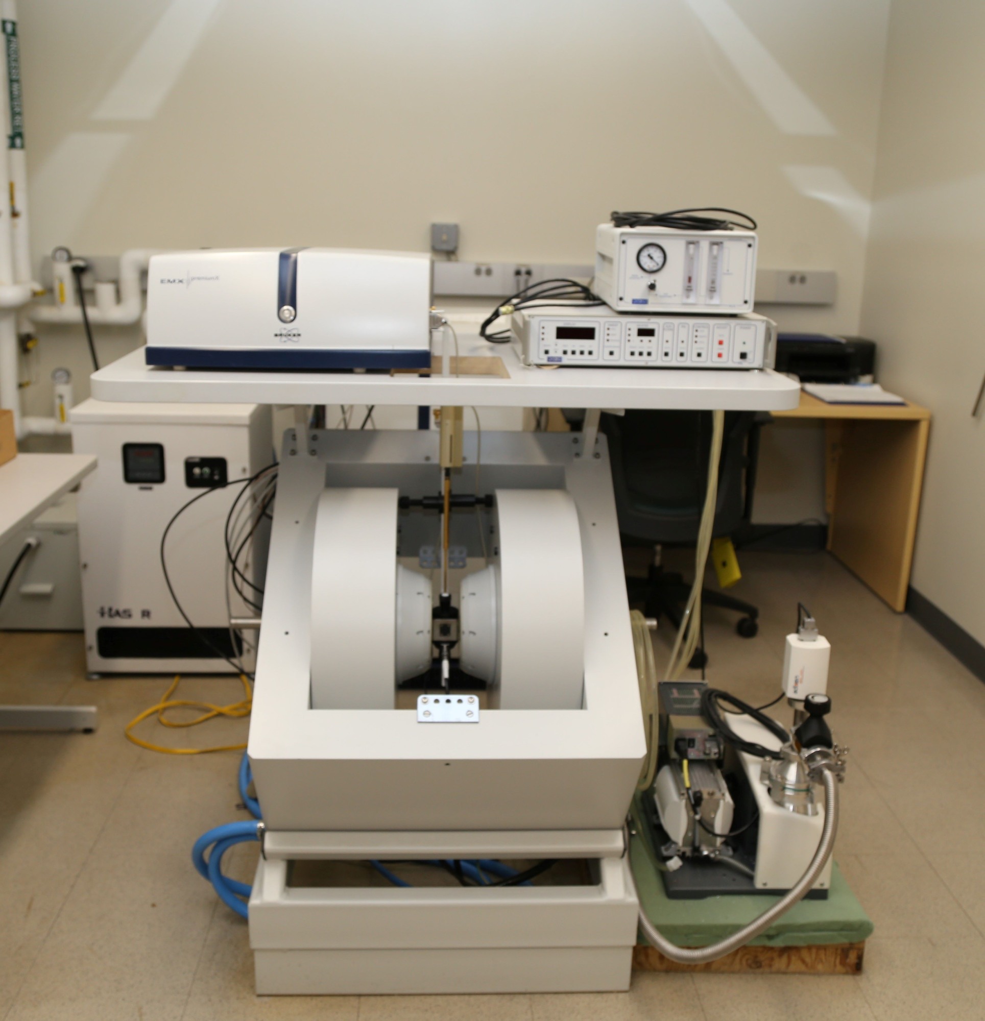
The Bruker EMX Electron Paramagnetic Resonance (EPR) Spectrometer is used for detection of paramagnetic species. The EMX EPR has a digital resolution of 24 bit center field, a field sweep up to 256,000 points, high-sensitivity resonator with irradiation window, dual channel, simultaneous detection of 1st and 2nd harmonic, and sensitivity S/N up to 2000:1. The system has a helium temperature control system for low temperature measurements in the temperature range 3.8 K (~-269 °C) to 300 K (~27°C). Applications span a wide range of areas an include kinetics of radical reactions, oxidation and reduction process, polymerization reactions, degradation of compounds by light, organometallic compounds and catalysis, free radicals, and antioxidants and radical scavengers.
Witec Alpha 300 Confocal Raman Microscope
The Witec Alpha 300 Confocal Raman Microscope is equipped with different scan modes for local Raman spectra, mapping distribution of phases within a sample and with 3D image generation and depth profile of samples. The depth profiling and 3D imaging capabilities of the Alpha 300 provide the ability to analyze the interior of transparent samples without microtome sectioning or freeze etching. Multi-wavelength couplers for selection of 457 nm, 532 nm, 633 nm, and 785 nm wavelength laser sources and a Ray Shield coupler for measurements of Raman spectra at low wave numbers. Fast Raman imaging with integration time in the milliseconds order per spectrum. A heating stage for in-situ measurements in the temperature range 25°C to 1400°C with a thermal stability of ± 0.3°C.