Electron Microscopy Facility Instrumentation
Focused Ion Beam (FIB)
FEI Helios 600 NanoLab Focuse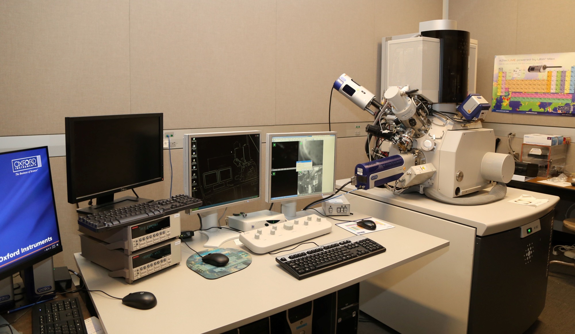 d Ion Beam (FIB) - Dual Beam (SEM)
This tool is fully equipped for (near) simultaneous ion/electron beam imaging. The Gallium ion beam can be used to remove material from the sample in user selectable patterns. Platinum (Pt) and Carbon are available for ion beam (or electron beam) stimulated deposition of conducting lines or areas. An Omniprobe AutoProbe 400 allows the physical manipulation of samples with nanometer precision and is used along with Pt metal deposition and standard FIB trenching to allow the selection and removal of electron transparent TEM samples from specific microstructural features. A NPGS pattern generation system (plus beam blanker and external scan interface) can control the electron beam for high resolution electron beam lithography. Composition analysis and mapping can be performed with the Oxford Aztec EDS system. Three dimensional crystallographic data can be generated from specimens using the fully automated Oxford EBSD system.
Scanning Electron Microscopy (SEM)
Thermo Scientific Quattro S - Environmental SEM 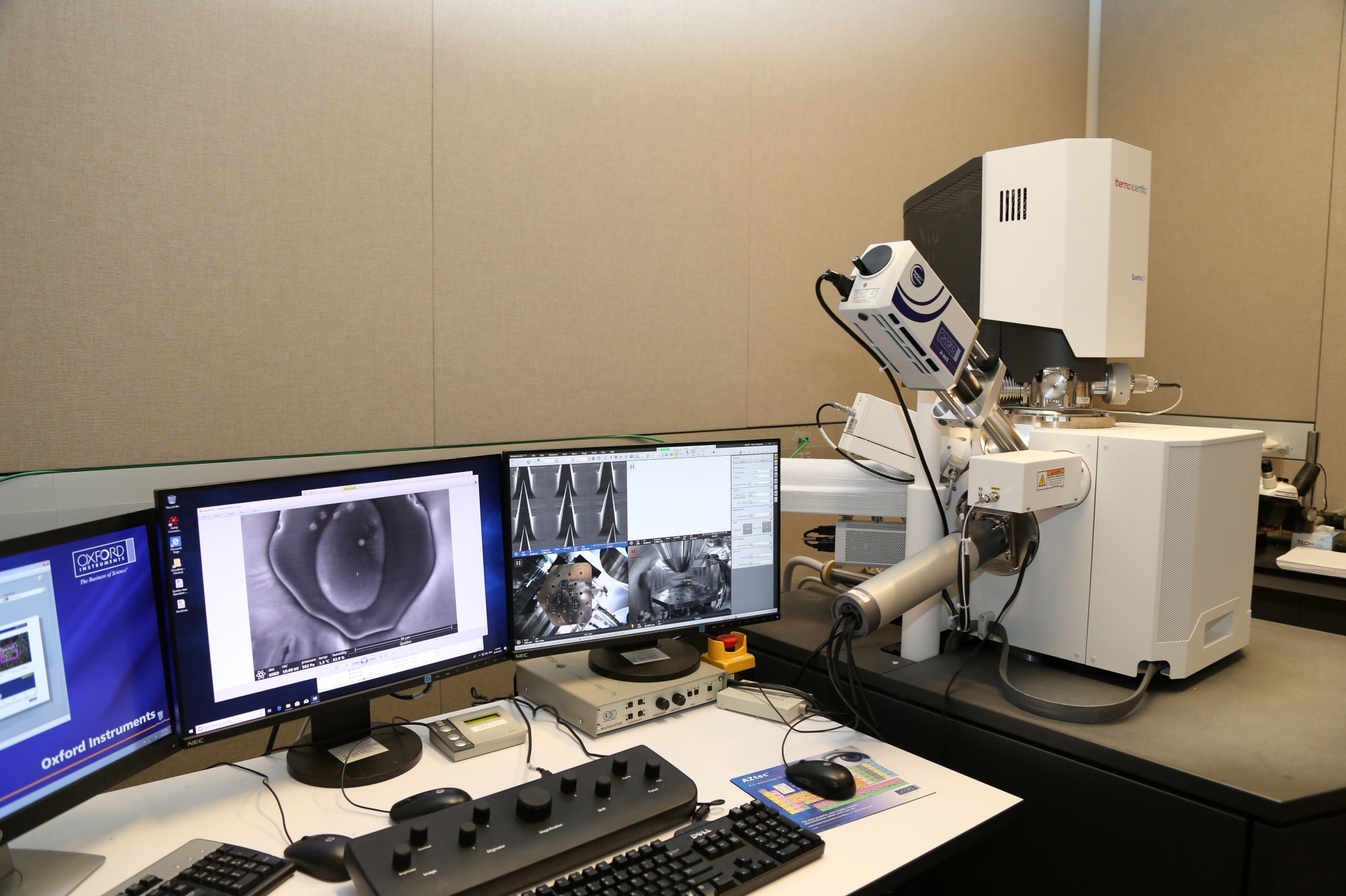
The Quattro environmental scanning electron microscope (SEM) is a unique SEM that combines a field-emission source for high-resolution imaging and an environmental feature allowing the observation of samples under wet/humid conditions. It is also equipped with an energy dispersive spectrometer (EDS) for analytical characterization and an electron backscatter detector (EBSD) for determining crystallographic orientations. The Quattro SEM can be operated in three different chamber pressure ranges: High Vacuum, Low Vacuum and Environmental.
The Quattro is also equipped with a cooling/heating stage. When cooled between 0 and 10 °C, specimens of very diverse nature can be imaged and analyzed at relative humidity conditions up to 100% (typical chamber pressures required are in the range 3-10 mbar). With the Cooling stage, humidity cycling experiments can be performed to characterize the sample's morphology and phase distribution at various relative humidity conditions. In-situ freeze-drying can be performed when the temperature range of the Cooling stage is set below 0 °C with a minimum of -20 °C. The Cooling stage can also heat the sample up to about 60 °C. The temperature and the ramp-up profile are directly controlled through the xT User Interface.
ThermoScientific APREO 2 SEM 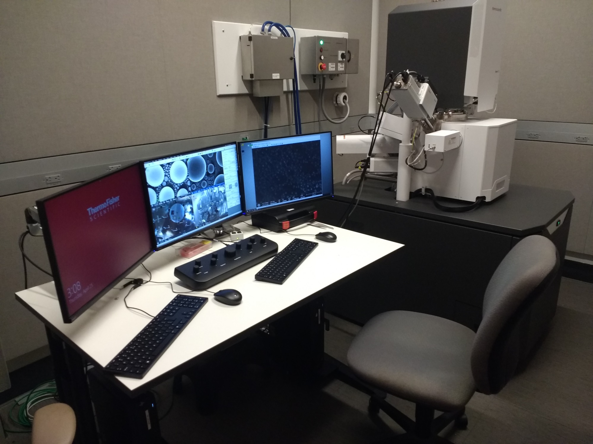
High Performance and Extreme Flexibility
The Apreo SEM has earned a reputation for its versatility and high- quality imaging performance- even on magnetic or other traditionally difficult samples. The Apreo 2 SEM improves its impressive resolution specifications and introduces live quantitative elemental mapping. A number of other new features are designed to make its advanced capabilities accessible for all users.
Highlights
• High performance, resolution, and contrast: The combination of advanced optics, detection, and automation in the Apreo 2 SEM makes high-resolution imaging possible even for users new to SEM
• Wide range of sample types: The Apreo 2 SEM is designed for maximum flexibility, with unique optics, detection, and beam control options that make highquality, high-resolution imaging possible regardless of target samples properties
• Live quantitative EDS: ChemiSEM Technology puts elemental information at your fingertips through live quantitative elemental mapping for unprecedented time to result and ease of use
• Quick and easy: Easy access to the stage, a convenient multi-sample holder, and fast pump down ensure no time is wasted loading samples; an automated routine to insert and remove a pressurelimiting aperture (PLA) allows for seamless switching between high and low vacuum without opening the chamber; and column presets allow you to save and reuse previously set parameters for easy access to the best imaging conditions
• Advanced automation: From optics to image acquisition, the Apreo 2 SEM provides a range of automated features to make imaging time as effective and efficient as possible; Flash Technology automates image fine tuning and the unique Undo function permits efficient exploration of imaging conditions; and Thermo Scientific Maps Software automates large area acquisition with up to four different simultaneous signals
• The Apreo 2 S includes a TEM grid row holder and STEM detector to do low voltage Scanning Transmission microscopy at much lower voltages than in traditional TEMs.
Transmission Electron Microscopy (TEM)
JEOL 2100F - S(TEM) 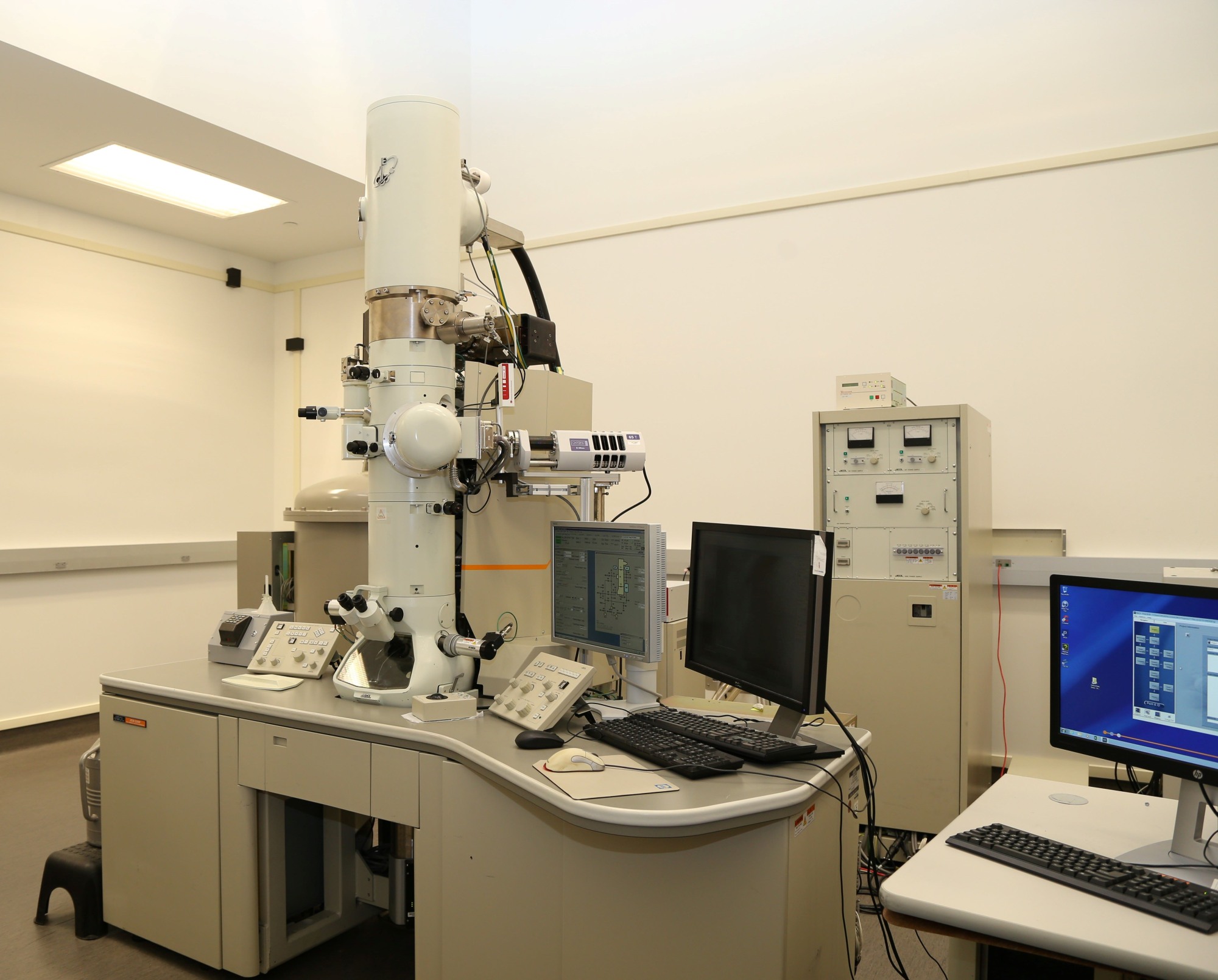
The 2100F is a 200 kV, field emission source, high-resolution/analytical S(TEM) capable of 0.10 nm lattice resolution. It is primarily used for obtaining lattice images or when chemical or crystallographic information is required from laterally small areas (1-10 nm). The instrument is equipped with an x-ray spectrometer capable of light element detection, a Gatan image filter (GIF) for element and phase analyses and energy filtered imaging, a high tilt holder for electron tomography and a high-resolution CCD camera system for digital image acquisition and processing. A low background double-tilt holder for chemical analysis by EDS, a standard double tilt holder, a heating holder capable of 1000C, and a heating straining holder are available. Probes as small as 1 nm in diameter are accessible for micro-diffraction experiments. STEM mode for EDS line and area scans, bright and dark field STEM detectors, and high angle annular dark field is capable in STEM mode.
Two in situ and operando specimen holders are now available for use on the 2100F. The MEMS Heating + Biasing Holder allows the user to perform heating (up to 1400 C), and simultaneous electrical-biasing, while observing the specimen in the TEM. The Single-Channel Gas Holder allows the user to perform TEM of specimens under atmospheric-pressure gas environment and heating (up to 1400 C). This expands our capabilities in the exciting area of in situ and operando TEM for studying phase transformations, battery materials/devices, solar materials/devices, electronic devices, catalysis, etc.
Philips CM 20 - TEM 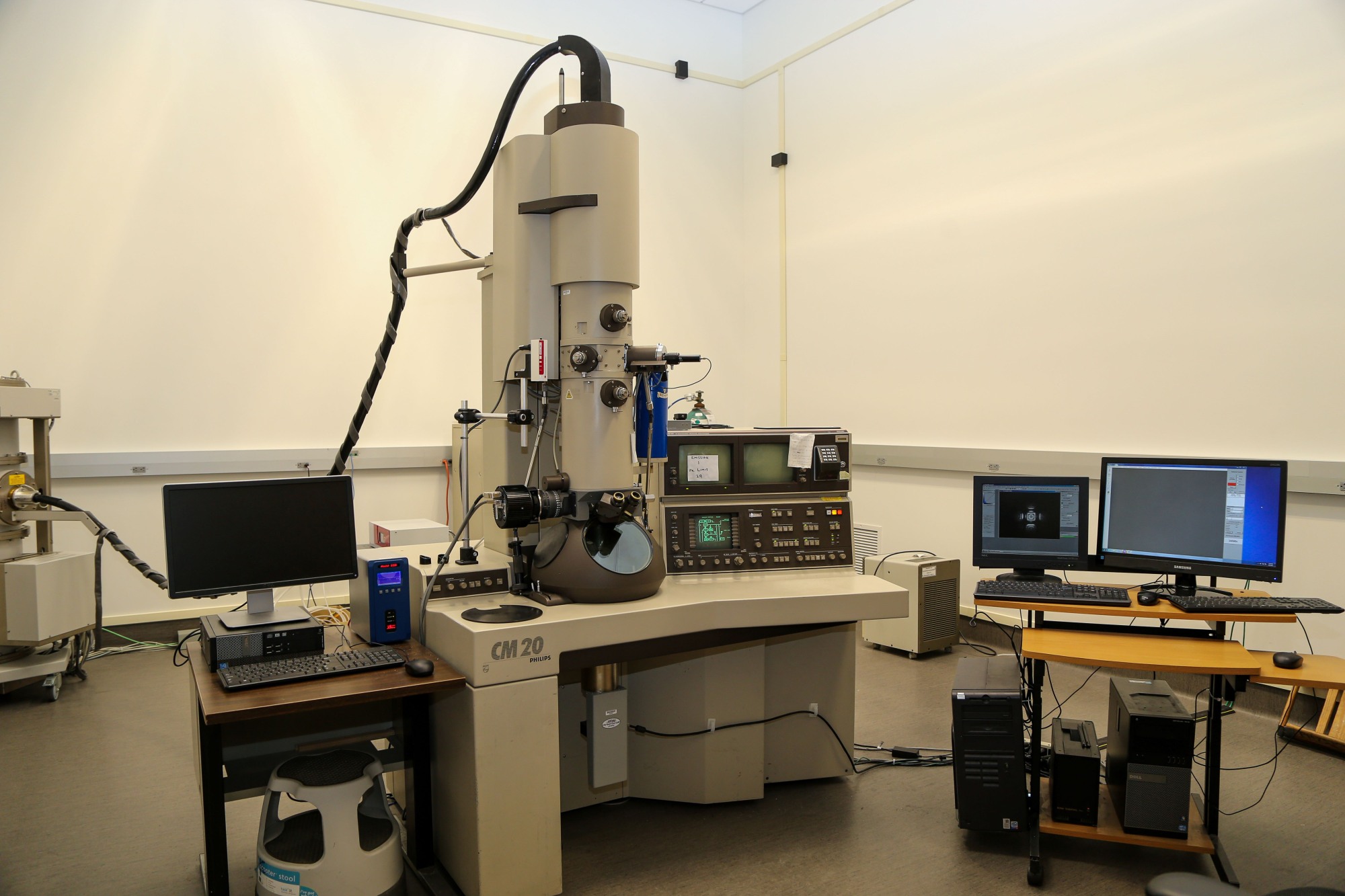
The FEI CM20 is available for applications that require high tilt, moderate resolution (0.23 nm). This 200 kV analytical TEM allows the large specimen tilts needed for dislocation characterization and weak-beam imaging applications. It is equipped with a low background double tilt holder and a liquid nitrogen temperature holder. Compositional analysis is performed on this instrument using energy dispersive x-ray spectroscopy. Images are recorded using a large field of view, side mounted, digital image acquisition system.
Surface Analysis
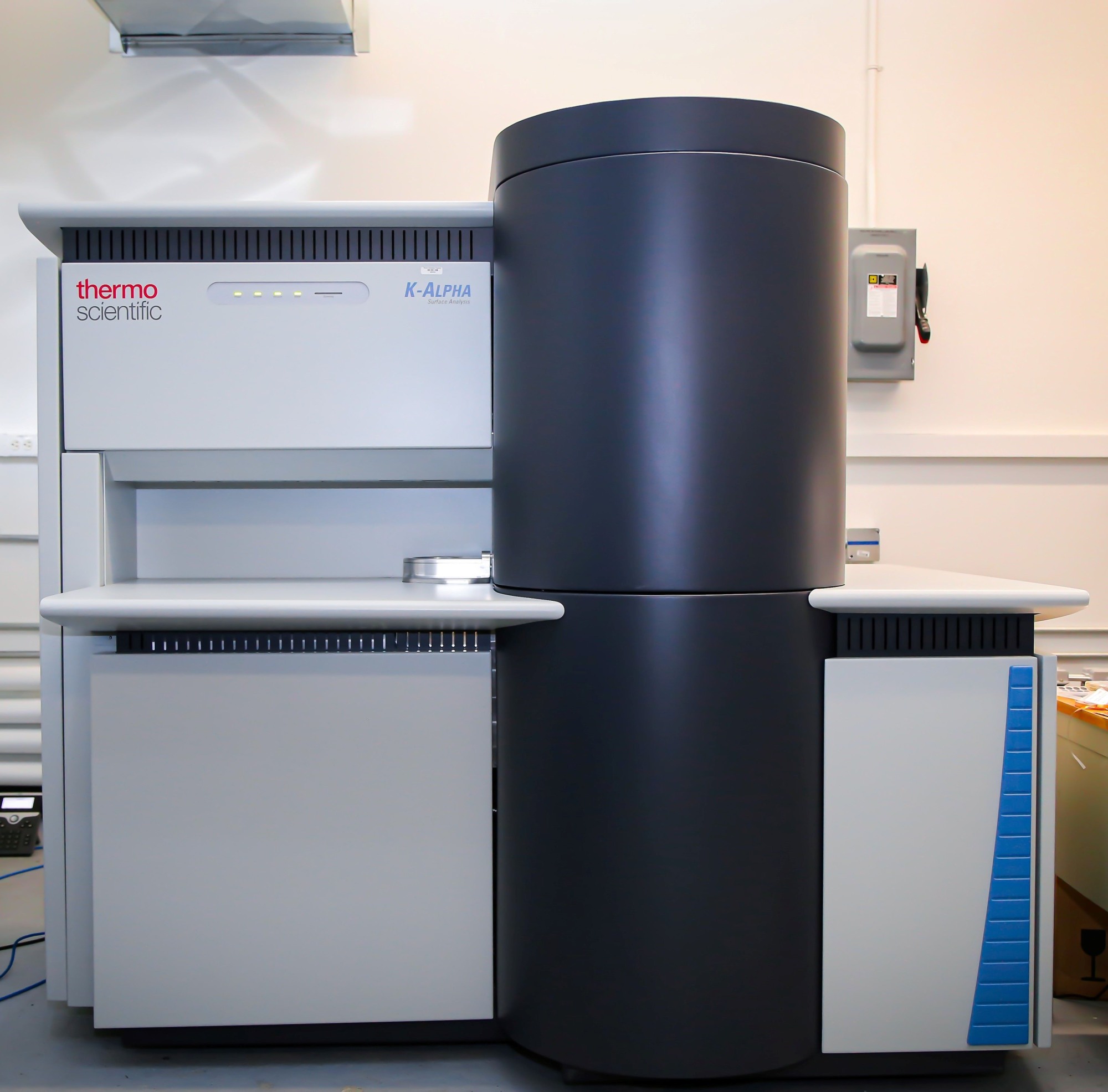
Thermo Scientific K-Alpha XPS
The K-Alpha X-ray photoelectron spectrometer (XPS) has a micro-focussed monochromated X-ray source, a double-focussing hemispherical analyzer with multi-element input lens (128-channel detector, 0-1486.29 eV range), and argon-ion source for depth-profiling/sample-cleaning. A transfer sample holder is available for samples sensitive to exposure to the atmosphere. Tilting and rotating sample holders are also available.
Sample Preparation Accessories
- Pd/Au Sputter Coater
- Carbon Thread Evaporator
- Plasma Cleaner
- Precision Ion Polishing System (PIPS)
- Ultrasonic Core Drill
- Dimple Polisher
- Tripod Polisher
- Low Speed Diamond Saw
- Low Speed Grinding/Polishing Wheels
- Vacuum Oven and Medium Temperature Oven
- Optical Microscopes